
In the Human Body
In the human body there are a total of about 1000 g of calcium , distributed:
- in bone tissue with structural function (99%);
- in muscle tissue (0.3%);
- in plasma , extracellular fluid and other cells (0.7%).
The calcium present in the plasma is represented, for 50%, by free calcium ions, for 40%, is bound to proteins and , for 10%, is complexed with anions. Among these three, the most important fraction is represented by ionized calcium (50%), since it is physiologically active, therefore rigorously controlled.
- it is necessary for the transmission of the nerve signal;
- is involved in the molecular mechanism of muscle contraction ;
- functions as an intracellular signal for some hormones , such as insulin ;
- it is necessary for the functioning of various enzymes thanks to which it intervenes, for example, in the coagulation cascade ;
- it is part of the intercellular cement that holds cells together at tight junctions;
Effects of hypocalcemia: tetany , cardiac hyperexcitability, bronchial , bladder , intestinal and vascular spasms .
Effects of hypercalcemia: reduction of muscle and nervous excitability.
To avoid the onset of these conditions, calcium levels are continuously kept under control thanks to the combined action of various hormones, such as calcitonin and parathyroid hormone.
Bone metabolism
Bone is a highly specialized connective tissue and, as such, composed of cells, fibers and amorphous ground substance. The latter, together with the fibers, constitutes the so-called extracellular matrix , formed in turn by a mineral component and an organic fraction.
The mineral component of the extracellular matrix is mainly formed by calcium phosphate , which organizes itself in the form of crystals, similar to needles, immersed in the organic component according to a precise orientation. The mineral component, also made up of phosphate, carbonate, magnesium , sodium and a small amount of water, represents only ¼ of the volume of the bone. However, being very dense, it alone constitutes half of the skeletal weight .
The organic component of the extracellular matrix, also called osteoid, is made up of collagen fibers (95%) and amorphous ground substance (5%), in turn composed of proteoglycans.
Bone is a dynamic structure, subjected to a remodeling process that continues throughout life. The extent of this process is considerable (about 1/5 of the skeleton is remodeled every 12 months) and, as such, requires a good supply of energy. Furthermore, to support bone remodeling , it is essential to associate the caloric intake with a good availability of minerals , especially calcium.
Responsible for bone renewal are two types of cells, respectively called osteoclasts and osteoblasts. The former, polynuclear and rich in microvilli, secrete acids and proteolytic enzymes which, by destroying the bone matrix, release the minerals it contains. Thanks to this process, approximately 500 mg of calcium is removed from bone daily (0.05% of total calcium). Following this process of bone erosion, osteoblasts intervene, cells with functions that are diametrically opposite to the previous ones. Osteoblasts, in fact, guarantee the formation and deposition of organic matrix in the cavities generated by the catabolic action of the osteoclasts. As soon as this matrix reaches a sufficient thickness, it is readily mineralized, thanks to the interposition of calcium. This mineralization process goes on for months, during which the density of the new bone progressively increases.
Most bone mass is built up by age 18-20; after this period the mineralization continues to increase, albeit slowly, until it reaches its peak around the age of thirty. For this reason it is very important to promote regular physical activity and adequate nutrition at a young age.
After the age of 40, bone mass undergoes a physiological reduction in terms of organic and mineral components. This process, absolutely physiological, therefore inevitable, is called senile osteoatrophy. Conversely, if the loss of bone mass is such as to compromise the performance of normal bone functions, we speak of osteoporosis . The difference between osteoatrophy and osteoporosis, therefore, is only quantitative. The two conditions, on the other hand, are the same from a qualitative point of view, because they share a reduction in bone mass due to the organic and mineral components.
Osteoporosis: Risk Factors
Many risk factors predispose to osteoporosis. Some of these are congenital and, as such, cannot be modified (female sex, white population, slender build , familiarity, age and menopause). For environmental or behavioral factors, on the other hand, a lot can be done:
- forced immobility (plaster of a limb, astronauts, etc.) there are specific therapies to accelerate bone remineralization);
- Diet low in calcium , Vitamin C (intervenes in the collagen maturation process) and Vitamin D (increases intestinal absorption of the mineral).
- Sedentary lifestyle (movement facilitates the deposition of calcium in the bones );
- Excess physical exercise (especially if not accompanied by an adequate intake of macro and micronutrients , it can accelerate bone decalcification );
- High protein diet ( too much protein promotes hypercalciuria, i.e. excessive elimination of calcium in the urine ); however, it should be noted that in various studies high-protein diets have been shown to increase intestinal calcium absorption, compensating for the increased urinary losses of the mineral; moreover, a diet very rich in proteins seems to favor the synthesis of hormones with anabolic effect on the bone (such as IGF-1 ), reducing the synthesis of parathormone ; at present, therefore, high protein diets are NOT considered harmful to bone health; even a low-protein diet, on the other hand, could represent a risk factor for osteoporosis.
- Abuse of alcohol and coffee
- Smoke
- Prolonged use of certain medications (such as cortisone )
The cessation of estrogen production increases the risk of osteoporosis in postmenopausal women , since the stimulatory effect of these hormones on osteoblastic proliferation is lost. Bone loss is particularly high in the first five years after the climacteric . Even in this delicate period of life, physical exercise has proved to be particularly effective in reducing the loss of bone mass.
| Recommended Intake Levels | |
| Age | mg/day |
| 0-1 | 500 |
| 1-6 | 800 |
| 7-10 | 1000 |
| 11-19 | 1200 |
| 20-29 | 1000 |
| 30-60 | 800 |
| >60 | 1000 |
| pregnancy and breastfeeding | +400 |
| for 5 years after menopause | 1500 |
Calcium and Vitamin D
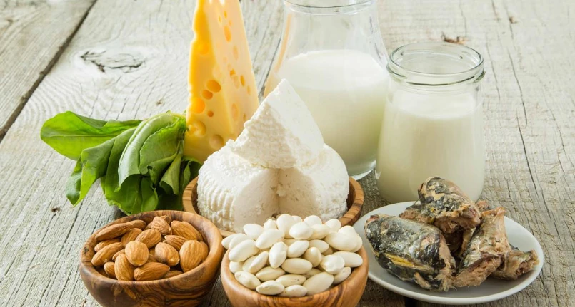
The presence of vitamin D is essential for the intestinal absorption of dietary calcium. This substance can be taken with certain foods ( liver , fish and fish oils , eggs , butter , milk and a few other foods ) or be synthesized in the skin .
7-dehydrocholesterol is formed from cholesterol which, by the action of UV rays on the skin , gives rise to vitamin D3. In turn, this vitamin must be activated, first passing through the liver , where it is hydroxylated, and finally through the kidneys , where it is completely activated. A vitamin D deficiency may therefore depend on insufficient food intake and/or insufficient exposure to sunlight . Furthermore, this deficiency may be linked to the presence of serious liver and/or kidney pathologies, which inhibit the activation of the vitamin.
Being fat soluble , vitamin D is stored in adipose tissue . This substance promotes intestinal absorption of calcium with the same mechanism as steroid hormones . As such, it enters the enterocyte nucleus and induces the coding for the synthesis of a protein, called calcium binding protein (CaBP) . This protein is capable of transporting calcium ions into enterocytes.
In essence, therefore, vitamin D is essential to increase the intestinal absorption of calcium taken with food. However, the amount of calcium ions that is absorbed also depends on other constituents of the diet . The bioavailability of calcium is in fact limited by the presence in the intestine of oxalates (contained in cocoa and green leafy vegetables such as spinach and chard), phytates ( bran , legumes , wholemeal bread ) and by the presence of too many lipids.
Given the importance of vitamin D for the intestinal absorption of calcium, its deficiency has repercussions in inadequate mineralization of the newly formed bone matrix. When this condition becomes chronic, it causes rickets in children and osteomalacia in adults.